Advantages And Disadvantages Of Laser Scanning Confocal Microscopes

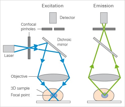
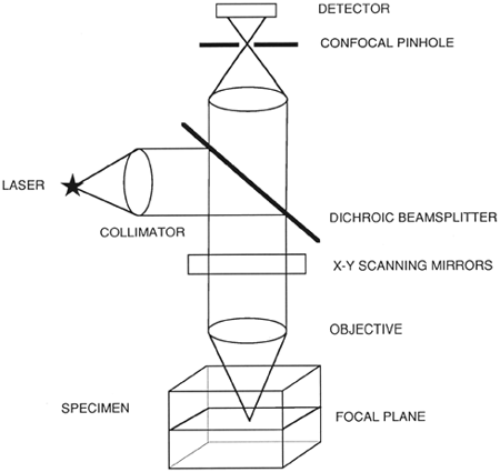
What Is Confocal Laser Scanning Microscopy
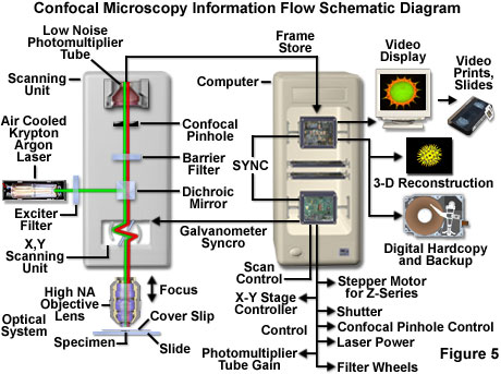
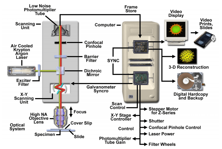
Confocal Microscopy Introduction Olympus Life Science

Confocal Microscope Principle Uses Parts Advantages And Disadvantages
Confocal Laser Scanning Microscopy Creative Biolabs
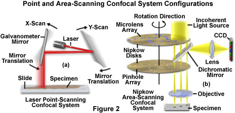
Confocal Microscopy Confocal Microscope Scanning Systems Olympus Life Science

Benefits Of Confocal Microscopy In Modern Life Science Applications Vision Blog
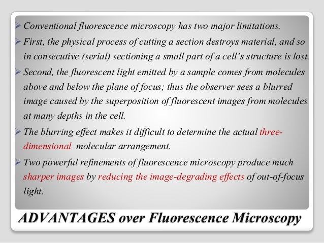
Advantages and disadvantages of confocal microscopy.
Advantages and disadvantages of laser scanning confocal microscopes.
Confocal Microscopy Principle Applications Ibidi

Confocal Laser Scanning Microscopy An Overview Sciencedirect Topics

Confocal Laser Scanning Microscopy An Overview Sciencedirect Topics
Confocal Laser Scanning Microscopy Clsm
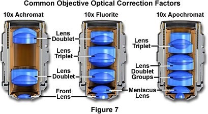
Confocal Microscopy Confocal Microscope Objectives Olympus Life Science
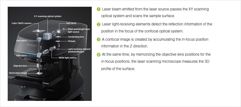
Profile Measuring Laser Microscopes Instruments Used For Roughness Measurements Introduction To Roughness Keyence America

Confocal Laser Scanning Microscope An Overview Sciencedirect Topics

Confocal Microscopy An Overview Sciencedirect Topics

Pdf The Basics Of Confocal Microscopy
How Does A Confocal Microscope Work
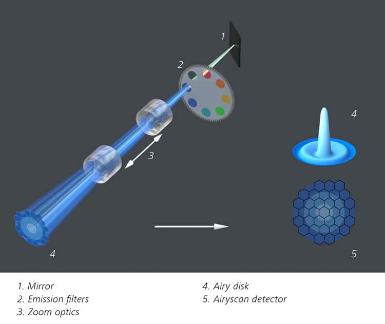
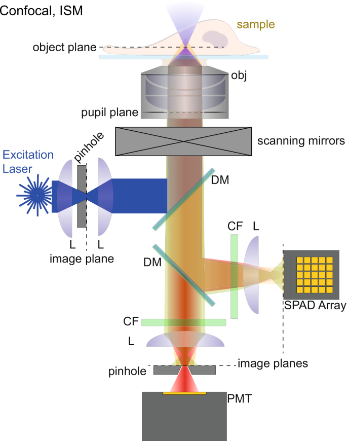
Zeiss Airyscan A Brave New Microscopy World With Sharper Confocal Resolution Bitesize Bio
Confocal Laser Scanning Microscopy Springerlink

Zeiss Microscopy Online Campus Introduction To Spinning Disk Microscopy
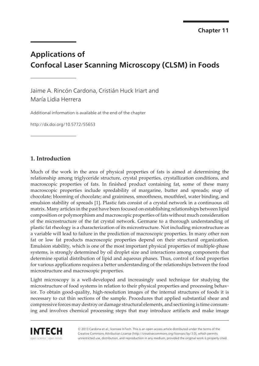
Pdf Applications Of Confocal Laser Scanning Microscopy Clsm In Foods

Confocal Laser Scanning Microscopy An Overview Sciencedirect Topics

Confocal Laser Scanning Microscope An Overview Sciencedirect Topics
Visualization Of Materials Using The Confocal Laser Scanning Microscopy Technique Chemical Society Reviews Rsc Publishing Doi 10 1039 C8cs00061a
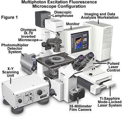
Multiphoton Fluorescence Microscopy Solutions Olympus Pour Les Sciences De La Vie
Https Encrypted Tbn0 Gstatic Com Images Q Tbn 3aand9gcr3fysoxor5w4y0kayjtt5nby84 Yhi3vdxn3rx2 E Usqp Cau

A Fluorescence In Situ Staining Method For Investigating Spores And Vegetative Cells Of Clostridia By Confocal Laser Scanning Microscopy And Structured Illuminated Microscopy Sciencedirect
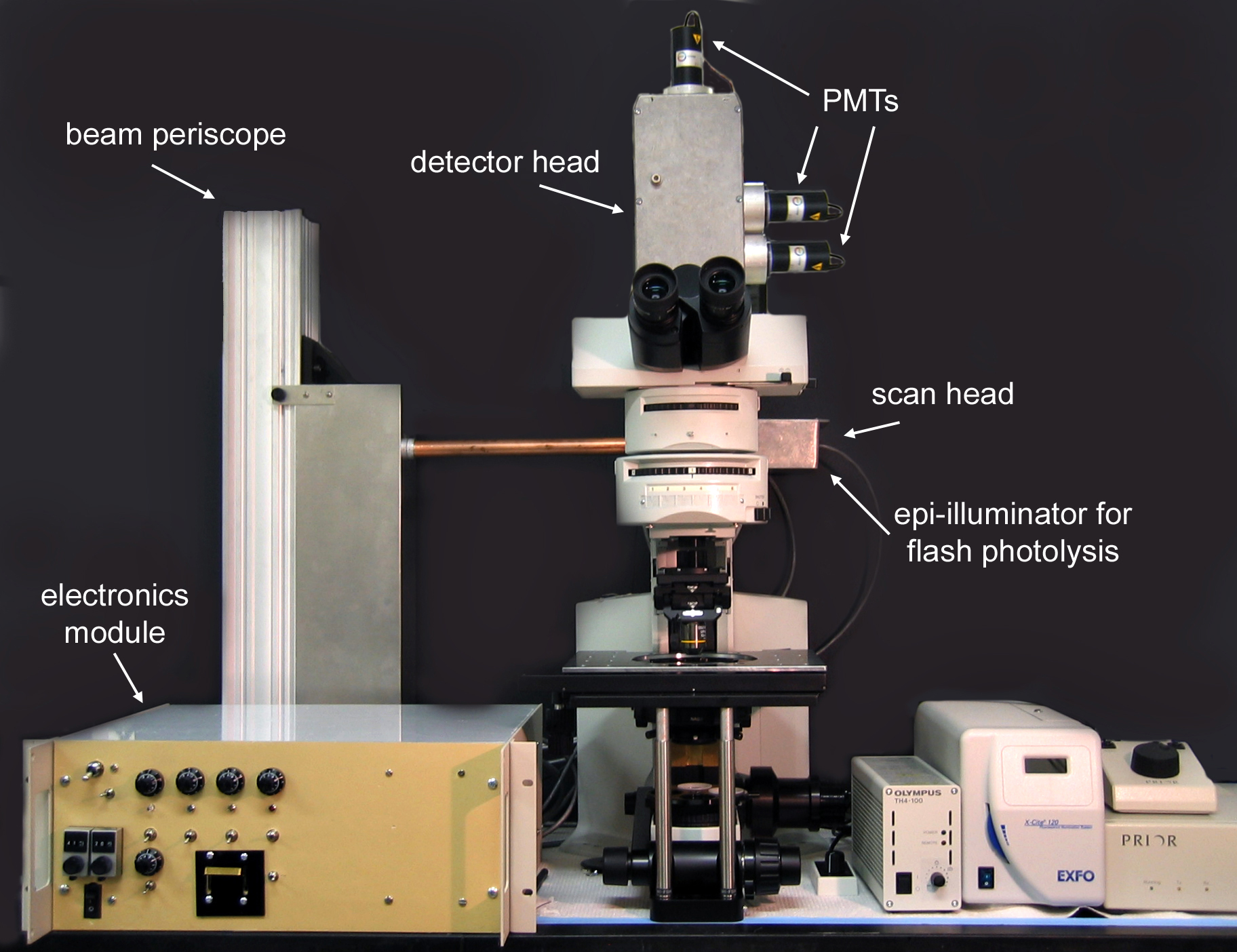
The Parker Lab At Uci Microscopy Construction How To Build Your Own Two Photon Microscope

Observing Cell Viability In 3d Cultures By A Confocal Laser Scanning Download Scientific Diagram

Confocal Microscopy Spectral Bleed Through Artifacts In Confocal Microscopy Olympus Life Science

Confocal Laser Scanning Microscopy An Overview Sciencedirect Topics
Source : pinterest.com
